|
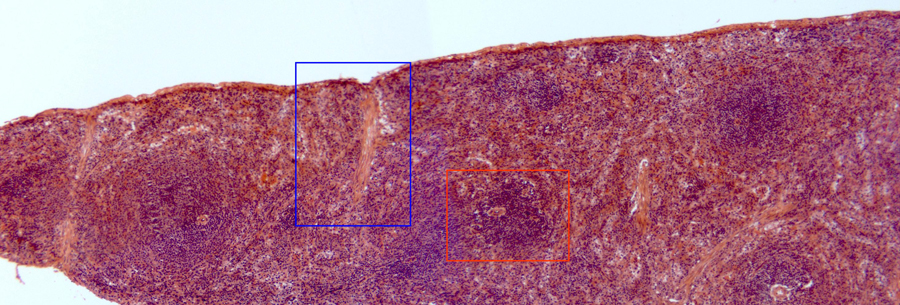
Photographic reconstruction of a spleen section stained with
hematoxylin/eosin
and observed with the objective of 10x. We can view the
capsule,
formed by densely arranged conjunctive tissue, and the spleeny parenchyma.
From the capsule leave
trabeculae
inward the organ. The spleeny parenchyma is formed by two zones:
white pulp
and
red pulp
.
The first one contains numerous lymphocytic components and the second one
includes a great amount of erythrocytes. Details of these structures can be
observed at higher magnification in other microphotographies (zones framed
in blue and red)
|