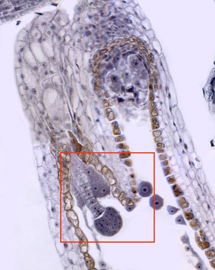
Aspect of a seminal rudiment observed with the objective of 40x. We can perceive the integuments and the nucellus around the embryo sac. Inside embryo sac we can see remains of endosperm and the embryo at early development stage. The embryo is united to the suspender, formed by a column of cells. At the end of the suspender is located a large basal cell. Laterally to the embryo are observed the antipodals cells. Details of the embryo are observed at higher magnification in other microphotography (zone framed in red)