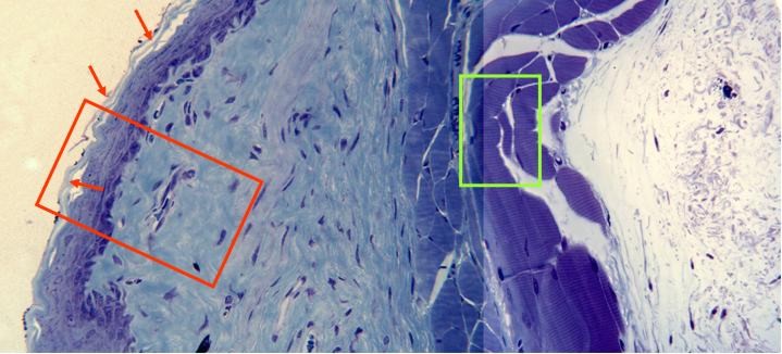
Photographic reconstruction of a
cross-section of the intestinal wall
observed with the objective of 40x.
Going from inside the
lumen
outwards we can see
the different layers that form the wall:
epithelium,
submucous
and muscular layer. The stratified
squamous
epithelium
can be observed,
while the outer layers are shown during moulting (arrows). The
submucous, of
loosely arranged connective tissue,
shows great amount of blue-stained
collagen fibers, numerous blood
vessels
and the cells
nuclei.
Finally, the
muscular
layer is formed, at this level, by
striated fibers and some blood
vessels.
In the microphotography we can also view
a longitudinally cut blood vessel
showing abundant red corpuscles inside.
Details of the epithelium and the
muscular layer appear at higher
magnification in other
microphotographies (zones framed in red
and green, respectively)