|
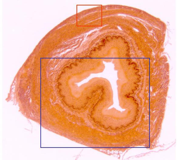
Complete
cross-section of esophagus stained with hematoxylin/eosin and observed with
the objective of 4x. Details of the different layers that form the esophagus
(zone framed in blue), as well as the muscular layer (zone framed in red)
can be observed at higher magnification in other microphotographies |