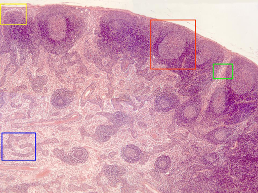
Photographic reconstruction of a lymphatic ganglion section
stained with hematoxylin/eosin and observed with the
objective of 10x. Going from outside inwards we can perceive
a conjunctive-type
capsule
that emits trabecula inwards. The
cortex
is located below the capsule and is formed by
lymphatic follicles.
Under the cortex is placed the
medulla,
which contains abundant blood
vessels
and is formed by
medullar-cords
and
-sines.
The follicles and the medullar cords are colored in blue and
show a dense aspect due to the presence of abundant
lymphocytes. Details of each structure are offered at higher
magnification in other microphotographies (zones framed in
different colors)