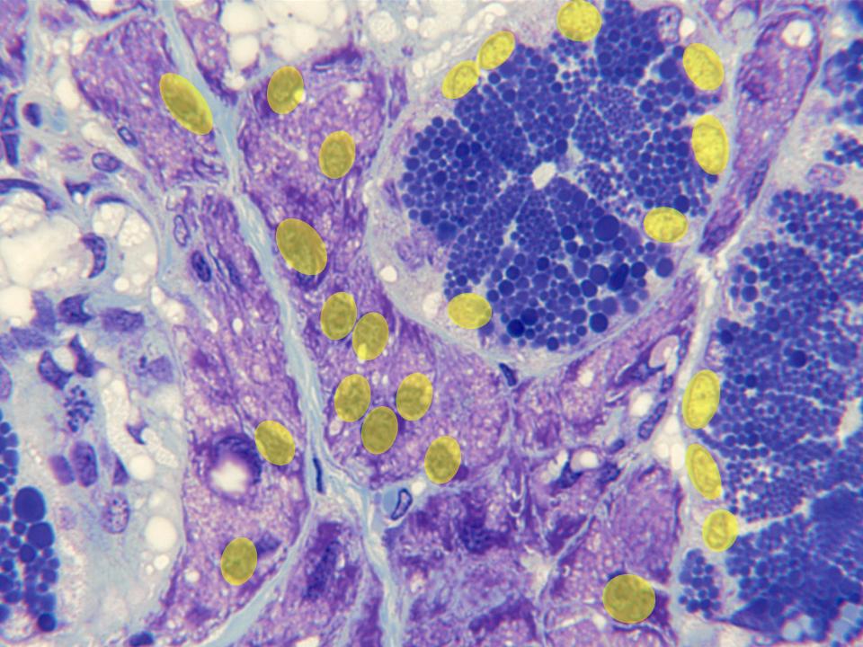
Detail of the acinar part of the
gland observed with the objective of
100x. We can distinguish the
serous
and
mucous acini,
as well as part of the network of
collagen septa
that separates them. The
nuclei (yellow)
of the secretory cells are visible
in the acini: they are stained in
dark blue in the mucous acini and in
light blue in the serous ones,
occupying the last ones a basal
position. The secreted material, in
form of small blue spheres, can also
be observed in the serous acini.