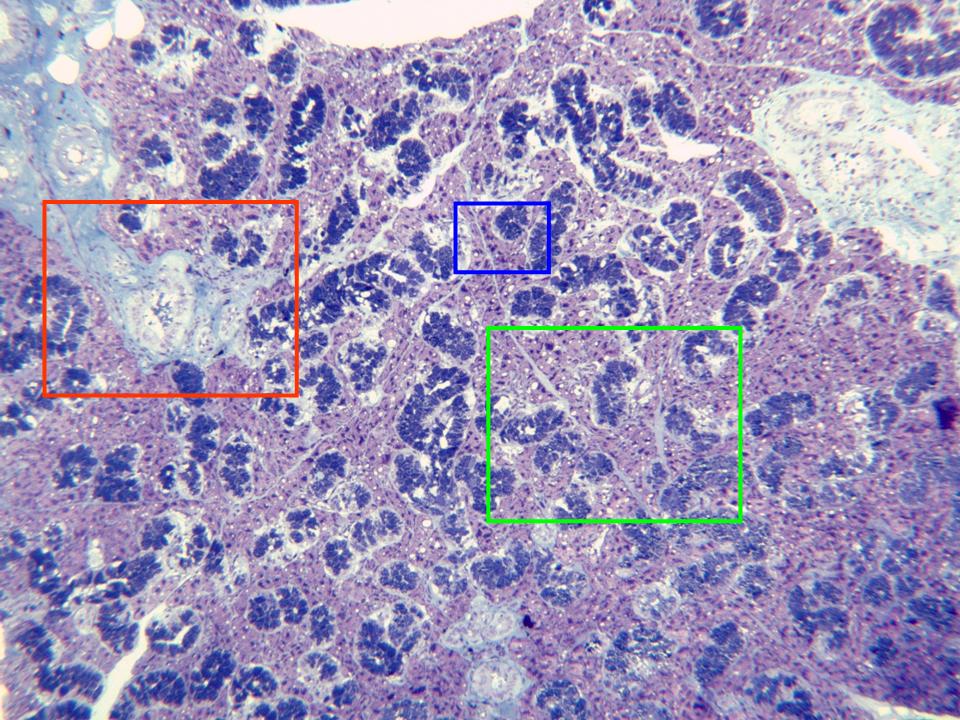
Semifine section of the gland stained with
toluidine blue and observed with the objective
of 10x. We can distinguish the different parts
of the gland: serous (dark blue-stained) and
mucous acini (bluish red-stained), separated by
fine collagen septa. We can also see the
excretory conduits
surrounded by connective tissue. Details of the
excretory conduits (box in red) and acinar zones
(boxes in blue and green) can be dobserved at
higher magnification in other microphotographies