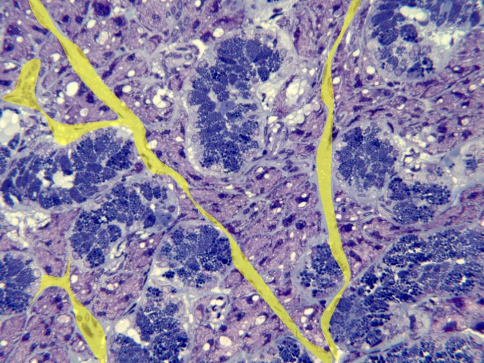
Aspect of the acinar part of the gland
observed with the objective of 40x. We
can identify the network of collagen
septa (yellow), separating the
blue-stained serous acini and
red-stained mucous acini.