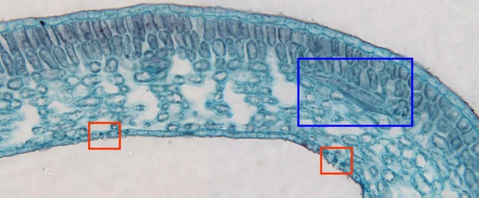
Photographic reconstruction of part of the mesophyll observed
with the objective of 40x. We can distinguish the upper and
lower
epidermis
and the
spongy
and
palisade parenchyma
(the last one shows large empty spaces between the cells)
containing a lot of chloroplasts peripherally located in the
cells. In the spongy parenchyma appears a longitudinally cut
vascular bundle,
visible at higher magnification in other microphotography (blue
box). In the lower epidermis are located some stomata (red
boxes), also visible at higher magnification in other
microphotography