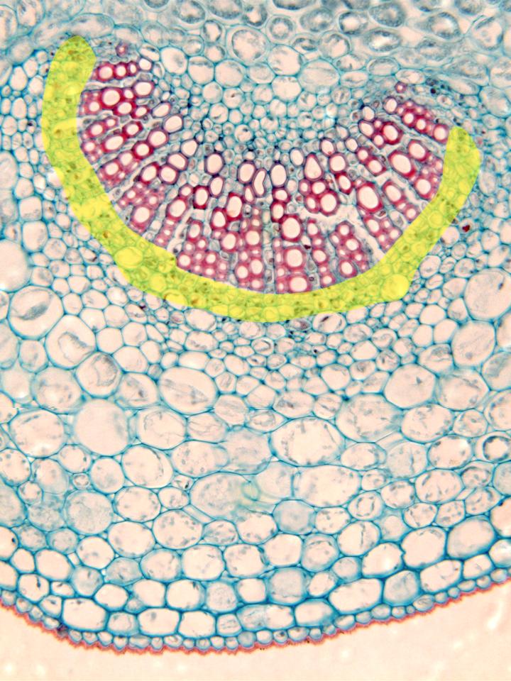
Photographic reconstruction of the zone corresponding to the central
nerve of the leaf observed with the objective of 40x. The image
shows the projection of the central nerve throughout the limb. The
most external part corresponds to the lower
epidermis,
whose cells are provided with a red-stained cuticle. Below it are
located the
collenchyma
(typical of this zone of the leaf) and the
assimilating parenchyma,
around the vascular bundle. This last element is formed by a part of
xylem
(located towards the zone of the foliar bundle) and a part of
phloem (yellow)
underneath. We can also perceive the vascular elements of the xylem,
showing the thick red-stained cell walls, radially arranged. The
parenchyma of the xylem is located in between, whose elements are
also radially arranged, but not red-stained