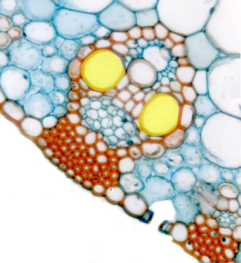
hotographic reconstruction of one of the vascular bundles of the leaf
observed with the objective of 100x. We can discern the
lower epidermis
, showing a
cuticle
on the cells and an open
stoma.
Under the epidermis appear two groups of
sclerenchyma fibers
(showing thick lignified cell walls), the bundle, and part of the
mesophyll
(formed by assimilating parenchyma). The vascular bundle, surrounded by
a
pod of sclerenchyma fibers,
contains conductive elements of
phloem
and xylem. The xylem is formed by two elements of
metaxylem
(yellow)
(of greater caliber) and other of
protoxylem
(of thinner caliber), next to which appears a
lysigenous cavity.
In the xylem are also visible some
sclerenchyma fibers
and the accompanying
parenchyma
around the lysigenous cavity
![]()