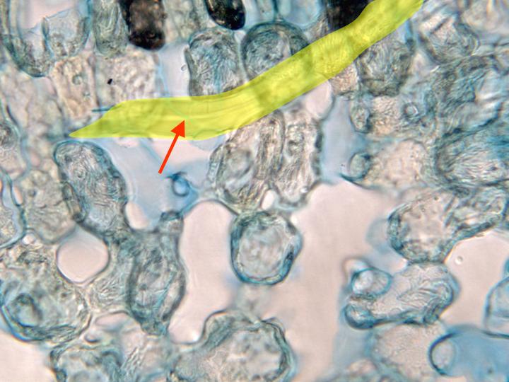
Aspect of the
spongy
parenchyma observed with the objective of 100x. The
image shows a longitudinally cut
sclereid (yellow)
(tricosclereid-type). We can notice the thickness of the
lignified secondary cell walls, leaving only a narrow
lumen in its interior. The cells of the
spongy and palisade
parenchyma show numerous acicular crystals inside