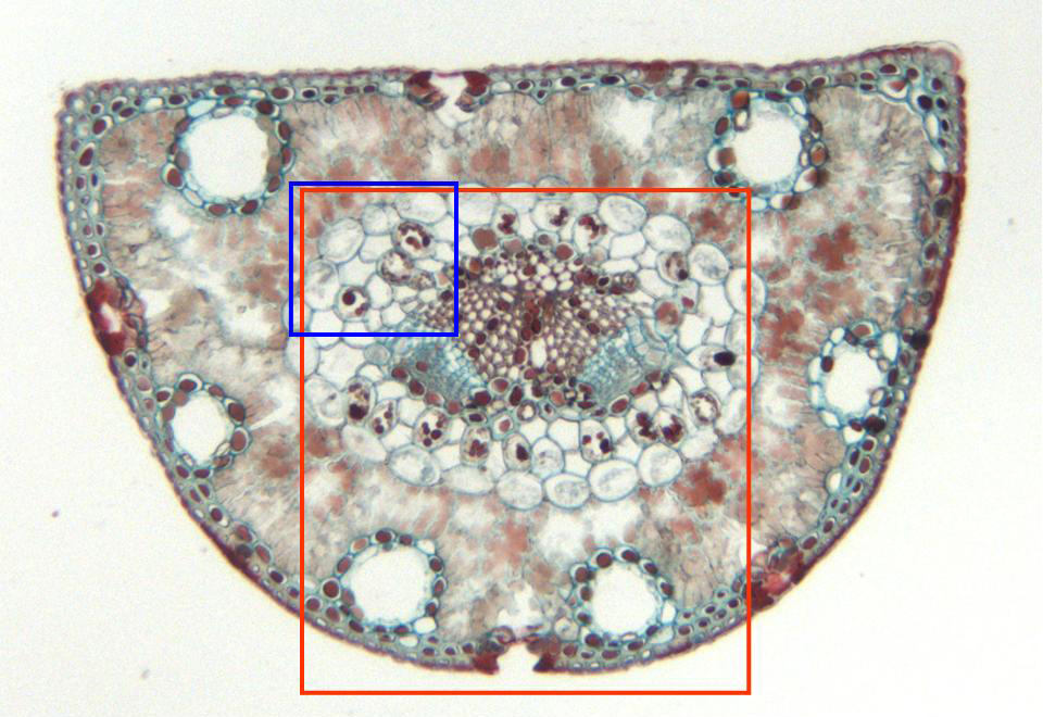
Cross-section of the leaf stained with safranin-light-green and
observed with the objective of 10x. We can perceive the
epidermis
(containing some
stomata),
the
mesophyll
and, below, two
vascular bundles.
In the mesophyll appear several transverselly cut
resin ducts.
Details of the vascular bundles and the surrounding tissue are seen
in other microphotographies (zones framed in red and blue)