|
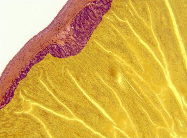
Aspect of the intestinal wall observed with the objective of 10x. Going from
outside inwards we can distinguish a thin serous layer or
adventitia,
the tunica muscular, integrated by two layers (external
and
internal),
the
submucous,
formed by connective tissue, and the mucous, containing some intestinal villi
(yellow).
Details of the intestinal villi can be observed in other microphotography
(zone framed in red). The
crypts of Lieberkühn
are located inside the villi, and they are provided with secretory cells,
which can be observed at higher magnification in other microphotography. |