|
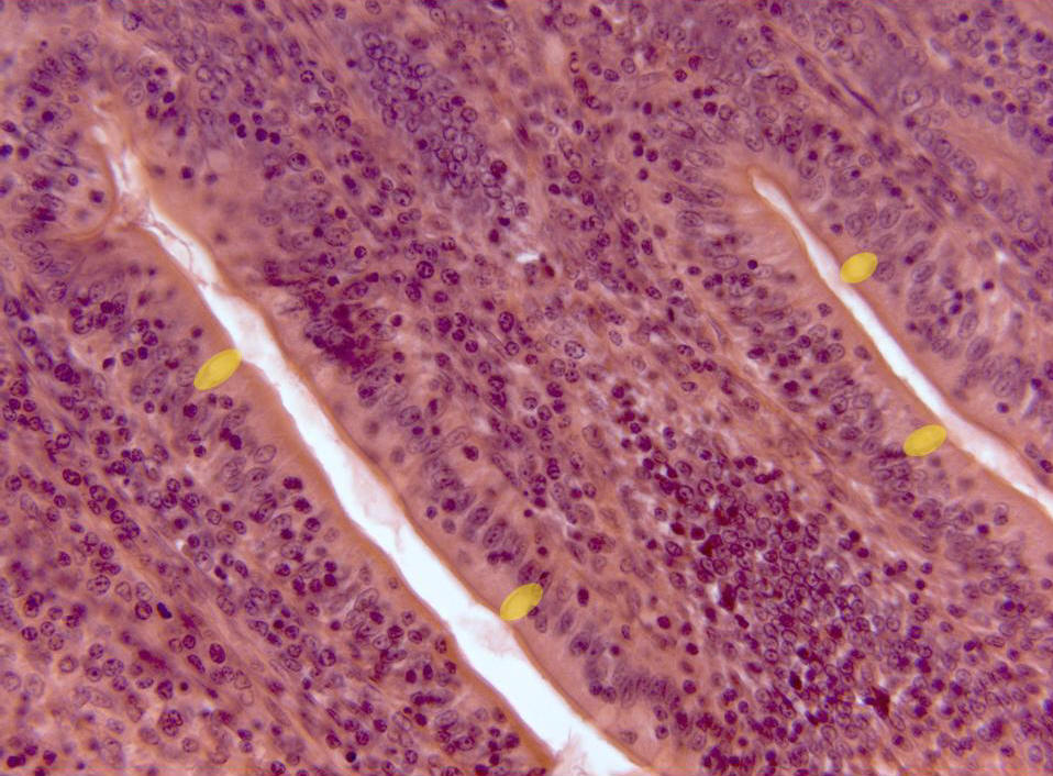
Detail of an
intestinal villus
observed with the objective of 40x. We can see the
intestinal lumen
in contact with the unistratified
epithelium,
formed by prismatic cells (provided with
glycocalyx)
and secretory cells (yellow).
Under the epithelium is placed the lamina propria, formed by
conjunctive tissue
and blood vessels, which can not be observed in this section. |