|
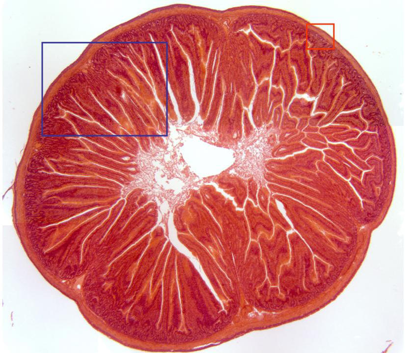
Cross-section of the small intestine stained with hematoxylin/eosin and
observed with the objective of 4x. We can observe the
lumen
in the center, as well as the
mucous,
the
muscular layer
and a thin
serous
layer externally. Details of these structures are observed at higher
magnification in other microphotographies (zones framed in red and blue) |