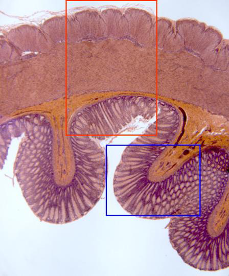
Section of the heavy intestine stained with hematoxylin/eosin
and observed and with the objective of 4x. Going from
outside inwards we can observe the different layers that
integrate the wall:
serous,
muscular, formed by the
external
and
internal layers,
submucous
and
mucous,
in contact with the
lumen.
Details of each zone are observed at higher magnification in
other microphotographies (areas framed in red and blue)