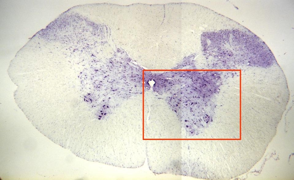
Photographic reconstruction of a spinal
cord section stained with cresyl violet
and observed with the objective of 4x.
We can observe the
gray matter,
formed by numerous neurons (in form of
H), and around it is located the
white matter.
Within the gray matter the neurons are
distributed in dorsal and ventral horns.
The
ependyma
can be seen in the center of the
microphotogray. Details of these
structures are offered at higher
magnification in other
microphotographies (zone framed in red)