This section offers the opportunity to examine the images (captured from the preparations) which form the Atlas, as real observations using the microscope. We can increase or diminish the observation field, as if different objectives were used, or to move freely within the preparation. It is recommended to use this option as a practice, once the user knows and has perfectly understood the detailed descriptions of each preparation. The images offered for exploration correspond to selected zones of the preparations, which have been photographed with the objective of 40x. The photographic reconstruction of all the area was made later (an image of which is shown next). Finally, by means of the software Zoomify, the web pages have been generated, allowing the complete exploration of them as it has been previously indicated.
ANIMAL ORGANS
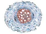
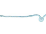
 Artery |
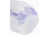
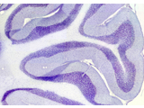 Cerebellum |
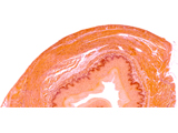
 Duodenum |
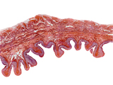
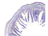
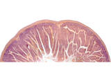
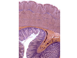
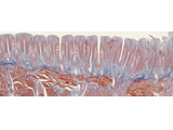
 Stomach |
 Compact bone |
||
 Spongy bone |
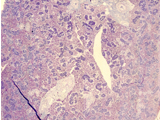
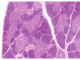
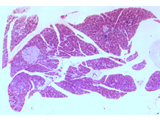 Pancreas |
 Trachea |
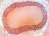
 Vein |
 Salivary gland
|
||
|
Spinal cord
|
Esophagus |
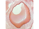
Urinary bladder |
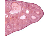
 Small intestine |
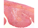
 Kidney
|
||
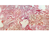
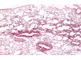 Lung
|
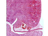
 Spleen
|
 Large intestine |
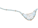
 Ovary |
 Cardiac muscle |
||
 Tongue
|
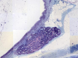
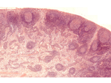 Lynphatic ganglion |
 Thymus |
||||
| VEGETAL ORGANS | ||||||
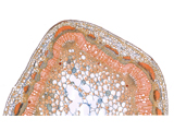
 Leaf of Pinus |
 Leaf of Nerium |
 Leaf of Jasminum |
 Leaf of Olea |
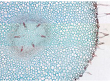
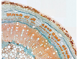
 Root of Zea |
||
|
Root of Vicia |
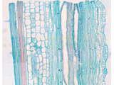
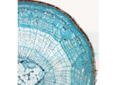
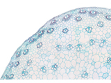
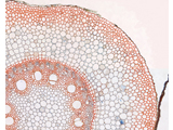
Stem of Cucurbita |
Stem of Hibiscus |
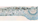
Stem of Tilia
|
Stem of Triticum |
||
 Stem of Helianthus |
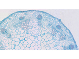
 Stem of Arachis (primary structure) |
 Stem of Arachis (secondary structure ) |
|
|
||
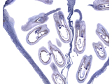
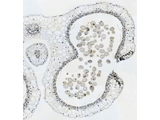 Anther of Lilium |
 Ovary of Lilium
|
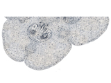
 Primordial embryo of Capsella |
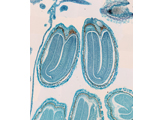 Mature embryo of Capsella |
 Epidermis leaf of Vicia |
||