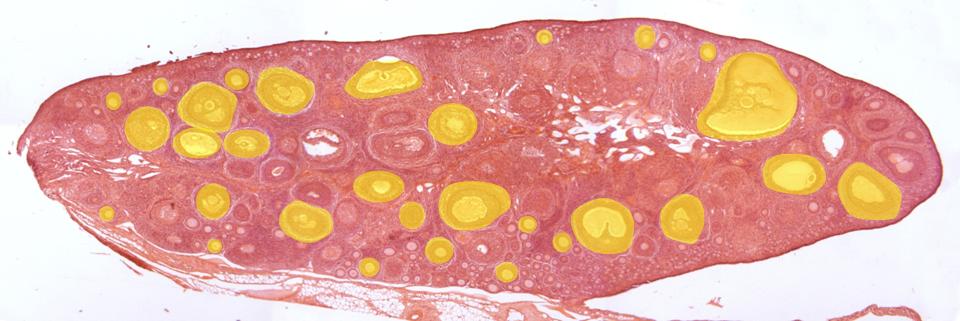
Photographic reconstruction of an ovary section stained with
hematoxylin/eosin and observed with the objective of 4x. We
can perceive the
epithelium
that covers it, as well as the
cortex,
containing many
follicles (yellow)
at different development stages, and a thin conjunctive-type
medullar portion,
internally located.