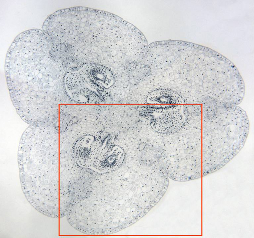
Complete cross-section of a tricarpelar ovary observed with the
objective of 4x. We can notice the
external
and
internal epidermis,
the
parenchyma
and the
seminal rudiments
within each ovarian cavity. We can also see the
vascular bundles,
located in the three carpelar leaves, easily identifiable with
polarized light. Details of the seminal rudiments are offered at
higher magnification in other microphotography (zone framed in
red)