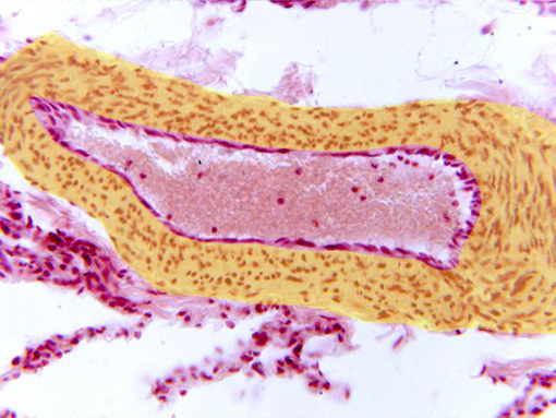
Detail of a small pulmonary venule observed with the
objective of 40x. The layers that integrate the wall are the
following:
intima
(formed by endothelium in contact with the lumen of the
venule),
media (yellow)
(showing the nuclei of the conjunctive cells), and part of
the
adventitia
(more externally located).
Around the vessel are visible some
interalveolar septa.