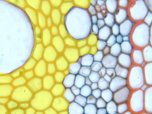
Aspect of the vascular cylinder observed with the objective of
100x. On the right of the microphotography appears part of the
endodermis,
whose cell walls show a thick lignification in form of U.
Underneath is located the
pericycle,
formed by a single layer of cells, surrounding the vascular
bundles of phloem and xylem. The xylem is represented by the
protoxylem
(more externally located and showing vessels of reduced
caliber), and the
metaxylem
(more internally located and showing large vessels). The
phloem
is placed next to the protoxylem, alternating with this one.
Around the xylem vessels are observed cells of lignified walls,
corresponding to the
parenchyma of the xylem and the sclerenchyma
(yellow)