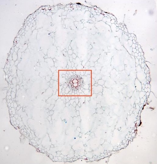
Aspect of a section stained with safranin-light-green and
observed with the objective of 10x. We can examine the
epidermis,
the
córtex
and the
vascular cylinder.
In the cortex we can distinguish an
external
and
internal
zone, differentiable by the cell size. Details of the
medullar zone can be observed at higher magnification in
other microphotography (rectangle in red)