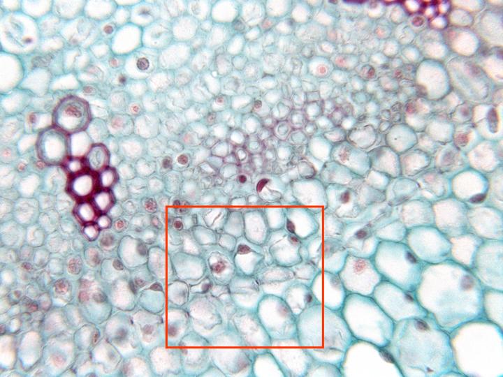
Microphotography of the medullar cylinder observed with
the objective of 40x. We can discern the vascular
bundles of
phloem
and xylem, surrounded by the
endodermis
and the
pericycle
(details of the zone framed in red can be observed at
higher magnification), as well as the
medullar parenchyma
and bundles of
sclerenchyma fibers.
Each xylem bundle is formed by a part of
protoxylem
(externally located) and a part of
metaxylem
(internally located), identifiable by the caliber and
position of the vessels. Part of the
cortical parenchyma
is also observed in the microphotography