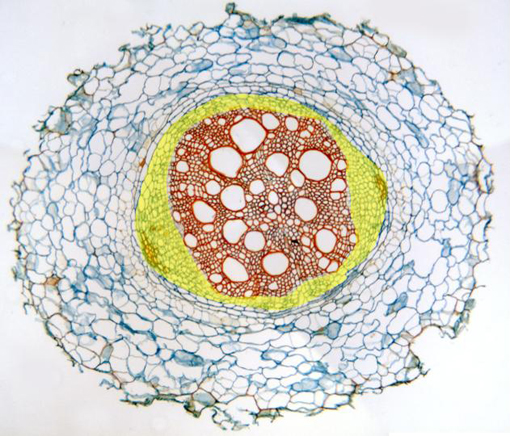
Complete cross-section of a root that initiates the secondary
growth stained with Alcian blue-safranin and observed with the
objective of 10x. We can view the
cortex
and the
vascular cylinder.
In the cylinder we can distinguish the
phloem
(yellow)
(externally located), showing two bundles of perivascular
sclerenchyma fibers,
and the
xylem
(internally located). In between are located the
endodermis and the pericycle,
visible at higher magnification in other microphotography. The
epidermis is missing, since the cells of suberin-deposited walls
have been removed