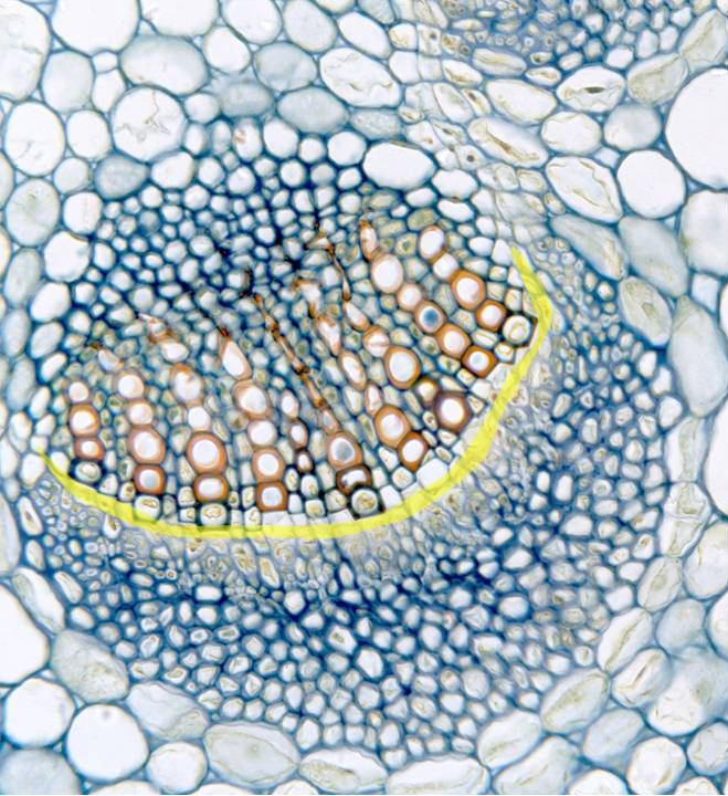
Aspect of a vascular bundle observed with the objective of 40x.
Going from outside (inferior part of the image) inwards (superior
part of the image) the stem are visible the following structures:
cortical parenchyma,
support tissue formed by
collenchyma,
showing blue-stained thick cellulose cell walls, vascular elements
of
phloem,
vascular cambium
(meristem), xylem, and
medullar parenchyma.
In the xylem, we can observe the
vascular elements
and the
xylematic radial parenchyma.
The cambium is formed by the
fascicular
part (yellow) (integrated in the vascular bundle) and the
interfascicular
part
(located between consecutive vascular bundles)