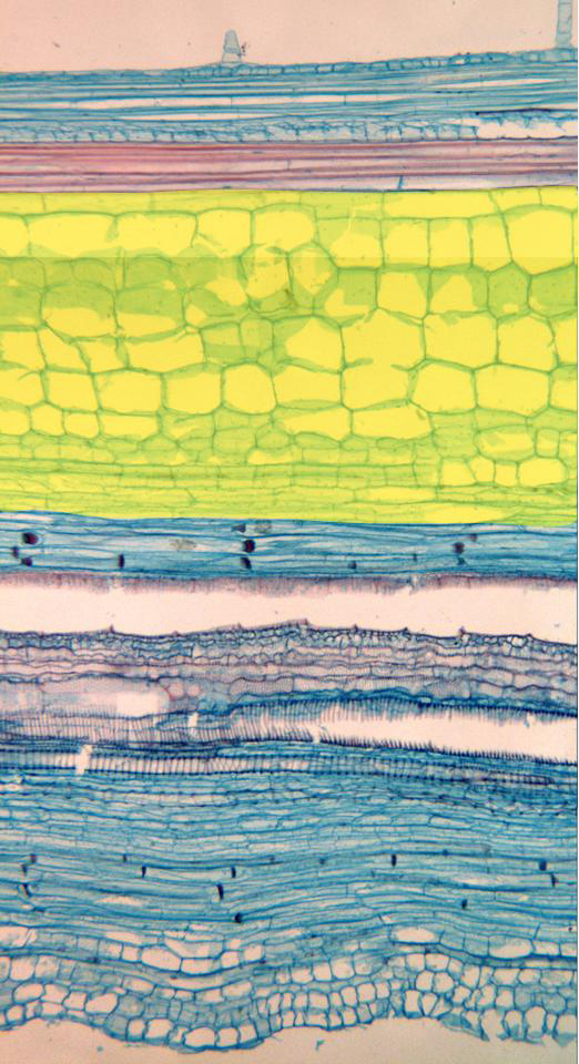
Photographic reconstruction of a longitudinal section of the stem stained with safranin-light-green and observed with the objective of 10x. Going from outside inwards (from top to bottom in the image) is shown the epidermis containing some trichomes, the subepidermal collenchyma, sclerenchyma fibers, the medullar parenchyma (yellow), phloem, xylem, and again phloem, since it is a bicollateral vascular bundle (with phloem towards outside and towards inside the xylem). We can discern sieve plates, located in the route of the vessels of the phloem
![]()