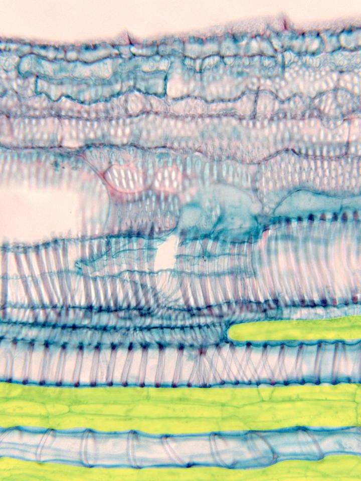
Aspect of a xylem bundle observed with the objective of 40x.
The cell walls of the vascular elements that form the bundle
are clearly visible due to the red-stained reinforcements of
lignin. More externally are placed the elements of the
metaxylem,
showing complicated reinforcements in the cell wall (it can
be easier observed in the microphotography observed with the
objective of 10x). More internally are located the elements
of the
protoxylem,
provided with reinforcements in form of ring or spiral.
Between these elements are placed cells of the
xylematic parenchyma (yellow)
![]()