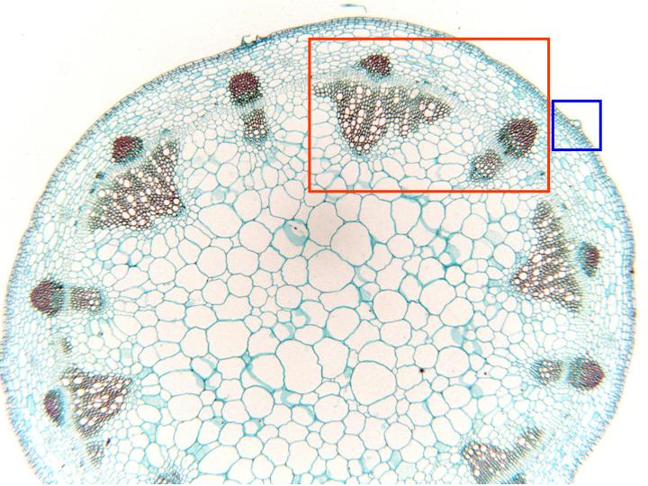
Cross-section of the stem stained with safranin-light-green and observed with the objective of 4x. We can see the different layers that form the stem: epidermis, cortex, vascular cylinder (constituted by the different vascular bundles), and the medullar parenchyma. Details of the vascular bundles and the epidermis are observed at higher magnification in other microphotographies (zones framed in red and blue, respectively)