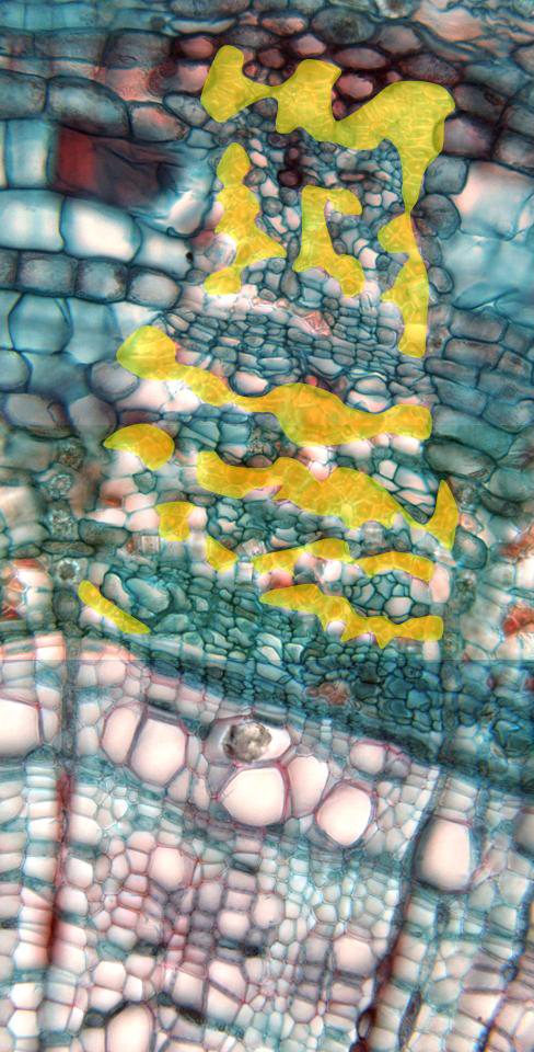
Photographic reconstruction of a vascular bundle observed with the
objective of 40x. We can view the vascular elements of the
phloem
(primary
and
secondary)
containing abundant intercalated
sclerenchyma fibers
(yellow),
as well as part of the elements of the
secondary xylem,
showing numerous parenchyma cells in between. Between the phloem and the
xylem we can see part of the
cambium. The
image also shows part of two
expanded phloem medullar rays,
whose cells contain crystalline inclusions in form of druse, as well as
some
medullar rays of the secondary xylem