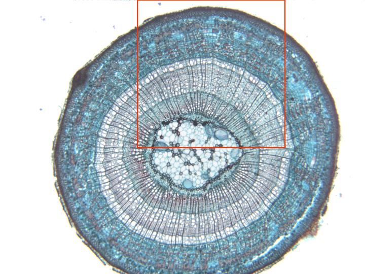
Complete section stained with safranin-light-green and
observed with the objective of 4x. Going from outside
inwards we can distinguish:
periderm,
cortex,
bundles of
phloem,
secondary xylem of the
first
and
second
year,
cambium,
and
medullar parenchyma.
Details of the cortex and the vascular zone can be observed
at higher magnification in other microphotography (box in
red)