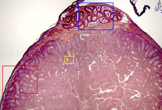
Photographic reconstruction of a testicle section stained
with hematoxylin/eosin and observed with the objective of
4x. We can see externally the
tunica albuginea,
while the interior is occupied by
seminiferous tubules.
In the superior part of the microphotography is observed a
portion of the
epididymis.
Details of tunica albuginea,
seminiferous
tubules and epididymis are offered at higher magnification
in other microphotographies (zones framed in blue, red, and
yellow)