 |
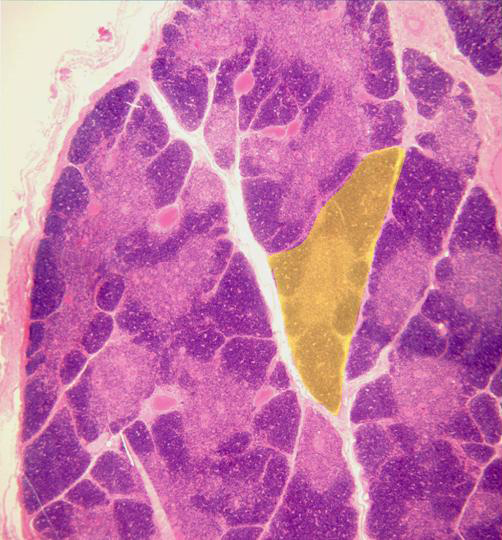
Photographic reconstruction of a thymus section stained with hematoxylin/
eosin and observed with the objective of 4x. We can observe externally the
thin conjunctive
capsule,
from which leaves septa towards the interior to divide the organ in numerous
lobules (yellow).
Each lobule shows an external zone with high cell content (cortex)
and other internal zone containing fewer cells (medulla).
|