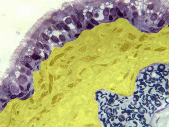
Detail of mucous and submucous observed with the
objective of 100x. We can observe the
pseudostratified prismatic
epithelium,
whose cells show large blue-stained nuclei and
abundant
cilia
in the apical pole. Under the epithelium is located
a
basal lamina
and, below, a
submucous
layer
(yellow)
formed by densely arranged connective tissue
(showing typical cells recognizable by the
blue-stained nuclei), bundles of
collagen fibers
and
blood vessels
containing
red corpuscles