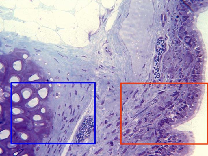
Semifine cross-section of trachea stained
with toluidine blue and observed with the
objective of 40x. In this image we can see
the different layers that constitute the
trachea:
mucous,
constituted by pseudostratified prismatic
epithelium, and
submucous,
formed by connective tissue containing
abundant blood vessels. We can also view
part of a
cartilage
and the external
adipose
tissue. Details of the mucous and the
cartilage are observed in other
microphotographies (zones framed in red and
blue, respectively)