|
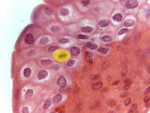
Aspect of the transitional
epithelium
observed with the objective of 100x. It is formed by several layers of cells
(the most internal layer shows some cells in stage of
mitosis [yellow]).
In contact with the epithelium is placed the conjunctive-type
lamina propria,
showing numerous fibrocytes with blue-stained nuclei
|