|
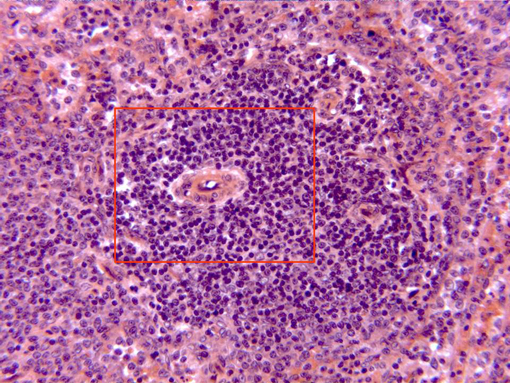
Detail of a
Malpighian corpuscle
observed with the objective of 40x. In the corpuscle we can see the
central artery,
surrounded by the
germinal center
and
containing many lymphocytes, and a transition
marginal zone
(the
red pulp
is
located on the left of this zone), containing fewer lymphocytes. Part of the
red pulp can be observed externally. Details of the central part of the
corpuscle are offered at higher magnification in other microphotography
(zone framed in red)
|