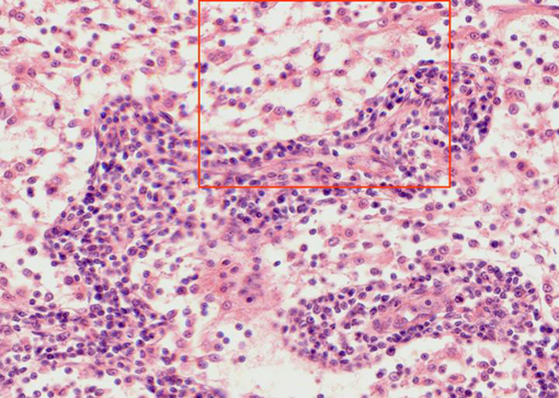
Aspect of the
medullar sines
observed with the objective of 40x. They are mainly formed
by reticular conjunctive tissue, showing the blue-stained
nuclei of the reticular cells. Two
medullar cords
can also be viewed, formed by many blue-stained lymphocytes.
The area framed in red appears at higher magnification in
other microphotography
