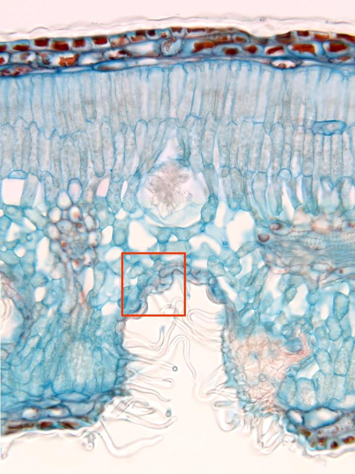
Photographic reconstruction of the complete thickness of the leaf
observed with the objective of 40x. From top to bottom we can
distinguish the following structures: externally is located the upper
epidermis
(whose cells show a thick translucent
cuticle),
next the
hypodermis
and, below, the mesophyll. The mesophyll is integrated by the
spongy
and
palisade parenchyma,
identifiable by the disposition and morphology of the cells, some of
which are
idioblasts,
containing crystalline groupings in form of druse. In the spongy
parenchyma are visible two
vascular bundles,
one of them in cross-section (surrounded by the
pod
of the bundle), and part of another cut longitudinally. The lower
epidermis shows invaginations or stomatal crypts, provided with stomata
(zone framed in red, detail in other microphotography) and covered by
numerous unicellular trichomes
![]()