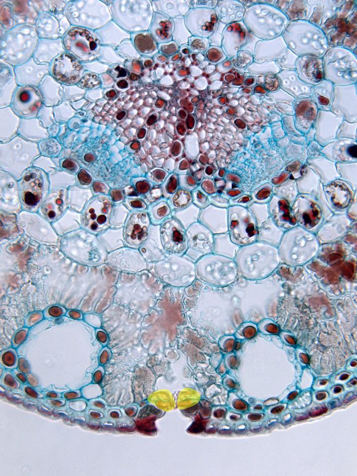
Photographic reconstruction of the vascular zone of the leaf and part of the mesophyll observed with the objective of 40x. Both vascular bundles are formed by a part of xylem and a part of phloem, surrounded by the transfusion tissue (formed by tracheids and parenchyma). These structures are separated from the mesophyll by a layer of cells denominated endodermis, of similar morphology and function to which contains the root. In the mesophyll are located two resin ducts, surrounded by cells of the assimilating parenchyma. The most external part is limited by an epidermis and, below, is placed the hypodermis. In the epidermis we can notice a stoma, showing the stomatal pore, the accessory cells and the guard cells (yellow), under which appears an empty space or substomatal cavity