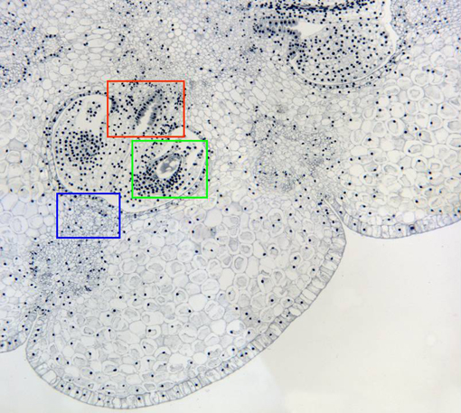
Aspect of part of the ovary observed with the objective of 10x.
The image shows the
external
and
internal epidermis,
the
parenchyma
in between, and three
seminal rudiments
within the ovarian cavities. Two of the
vascular bundles
are also visible. Details of the placenta, a seminal rudiment
and a vascular bundle are observed at higher magnification in
other microphotographies (zones framed in red, green, and blue,
respectively)
