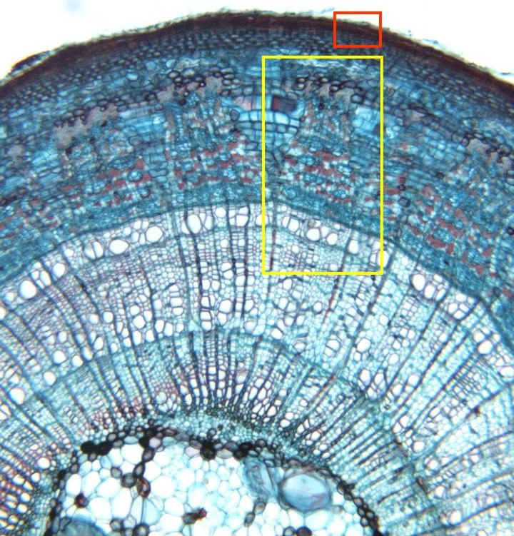
Photographic reconstruction of the epidermis, the
cortex
and the vascular cylinder observed with the objective of 10x. We can see
the
periderm,
details of which appear in other microphotography (box in red),
phloem,
details of which are offered in other microphotography (box in yellow),
primary xylem,
secondary xylem of the first year,
secondary xylem of the second year,
xylem medullar rays
crossing the thickness of the xylem,
expanded phloem medullar rays,
cambium,
and
medullar parenchyma