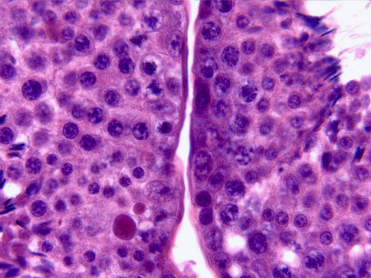
Detail of the wall of two contiguous
seminiferous
tubules observed with the objective of 100x. We can perceive
the wall structure, constituted by germinal cells at
different development stages and by
Sertoli
cells. Going from outside inwards we can distinguish
different types of germinal cells:
spermatogonias,
primary
and
secondary spermatocytes
and
spermatids
(they are
more developed
when nearest to the tubule
lumen)
. Each tubule is surrounded by a thin
lamina propria
and between the tubules there is
connective tissue.
![]()