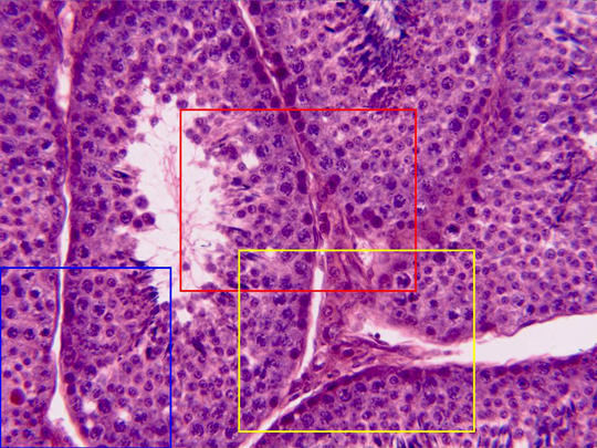
Aspect of the interior of the testicle observed with the
objective of 40x. It is totality occupied by
seminiferous tubules
separated by
connective tissue.
We can see a group of
interstitial cells,
as well as the wall thickness and the
lumen
of a tubule. Details of the tubules wall (zones framed in
red and blue) and the interstitial cells (zone framed in
yellow) are observed at higher magnification in other
microphotographies
![]()