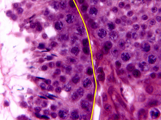
Detail of the wall of two contiguous
seminiferous tubules
observed with the objective of 100x. In the wall, formed by
germinative epithelium (already described in other
microphotography), we can notice
Sertoli cells,
spermatogonias,
primary spermatocytes
and spermatids at
major
or
minor
development stage (the more developed are close to the
lumen).
Each
seminiferous
tubule is surrounded by a
lamina propria (yellow),
in which we can distinguish some
miofibroblasts nuclei. 