|
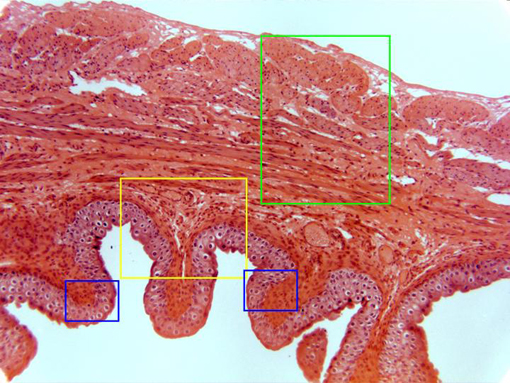
Cross-section of the vesical wall stained with hematoxylin/eosin and
observed with the objective of 10x. At this magnification we can see the
adventitia
layer (more externally located), the
longitudinal
and
circular
muscular layers,
the
lamina propria,
and the
epithelium
(in contact with the
lumen)
showing a number of folds. Details of these layers are observed at higher
magnification in other microphotographies (zones framed in blue, yellow, and
green) |