|
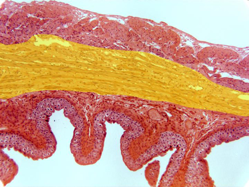
Cross-section of the vesical wall stained with hematoxylin/eosin and
observed with the objective of 10x. At this magnification we can see the
adventitia
layer (more externally located), the
longitudinal
and circular
(yellow)
muscular layers,
the
lamina propria,
and the
epithelium
(in contact with the
lumen)
showing a number of folds.
|