![]()
 |
|
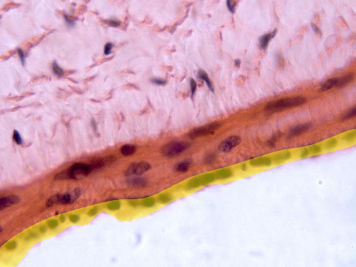
Details of the
three layers of the vein wall can be
observed with the objective of 100x: intima
(yellow showing
the nuclei of the endothelial cells),
media (showing
the nuclei of
the smooth muscle fibers), and
adventitia
(showing the
nuclei
of the fibrocytes and the
collagen fibers)
|