![]()
 |
|
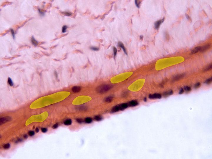
Details of the
three layers of the vein wall can be
observed with the objective of 100x:
intima (showing
the nuclei of the endothelial cells),
media (showing
the nuclei (yellow) of
the smooth muscle fibers), and
adventitia
(showing the
nuclei
of the fibrocytes and the
collagen fibers)
Details of the three layers of the vein wall can be observed with the objective of 100x: intima (showing the nuclei of the endothelial cells), media, showing the of the smooth muscle fibers, and adventitia (showing the nuclei of the fibrocytes and the collagen fibers) |